Ultra-Small Bacteria
May 7, 2015
In my early
audiophile days, bigger was always better. Since the best
magnets of the time were
Alnico, with
magnetic strength far less than
today's magnets,
loudspeakers were
inefficient. Many
watts of
power were required to drive these, and this meant large
amplifiers. Coupling from the
high impedance outputs of
vacuum tubes to the low impedance loudspeakers was done by
transformers, and these transformers were built with large, heavy
cores to preserve low
frequency response.
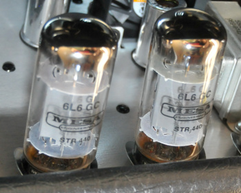
The 6L6 was the most popular type of vacuum tube in the output stage of power amplifiers.
The 6L6 is still used today in some guitar amplifiers.
(Photograph (cropped) by Art Brom, via Wikimedia Commons.)
Today's trend is towards smaller
electronics with more functionality. The minimum feature dimension in
transistors has reached
14 nanometers, and there are
plans in place to bring this down to 5 nm in 2020. Since the distance between
atoms in
silicon is about half a
nanometer, a five nanometer
field effect transistor channel would encompass just ten atoms of silicon.
There are hopes that new
materials such as
graphene will aid in this "race to the bottom," but we can't help but think that a limit will be reached. While
science fiction fans wait for
positronic brains,
computer scientists have been improving
computational speed using
multiple processors. It's likely that the device with which you're reading this article operates by dividing computing tasks among severtal processors.

Image of the die for the Advanced Micro Devices, Inc. quad core Opteron processor.
Notice the symmetry about a central point.
(Image ©2008 Advanced Micro Devices, Inc. with permissions for reuse, via Wikimedia Commons.)
Just as there's an apparent limit to how small a transistor can be, there's also a limit to how small a
living cell can be. In assessing this limit, we must immediately eliminate
viruses, which might appear to be the smallest living things. Viruses, however, are not alive. They're just pieces of
RNA or
DNA protected by a
protein sheath. Since they have no inherent ability to
replicate on their own, instead relying on the
reproductive mechanisms of living cells to make copies of themselves, they do not meet the definition of "living."
While the size of the smallest cell is much larger than that of a transistor, they are difficult to image accurately, since high resolution imaging devices require a
vacuum, and vacuum will modify or destroy the cell structure. A team of
scientists from the
University of California (Berkeley, California),
Lawrence Berkeley National Laboratory (Berkeley, California), and the
Joint Genome Institute (Walnut Creek, California), have developed techniques to accurately image small cells, and they applied their techniques to ultra-small bacteria cells in
groundwater.[1-2]
Since imaging ultra-small bacteria has been difficult, there has been no consensus on the range of sizes for bacteria. This new study is the first comprehensive
electron microscopy and
genomic study of ultra-small bacteria.[2] As a reference to the size of these cells, which have an average
volume of just 0.009 cubic
micrometers, 150 of them can fit inside a cell of the typical bacterium,
Escherichia coli, and 150,000 such cells would be needed to cover the tip of a
human hair.[1-2]
These ultra-small bacteria were collected by repeated
filtering of groundwater, collected at
Rifle, Colorado, through filters down to 200 nanometers. The last filter size is typically used to
sterilize water, but the presence of these bacteria showed that the water was not really sterile.[2] The bacteria were
flash frozen to -272 °C using an unique device called a cryo plunger that ensured that they weren't altered in transit from the field to the
laboratory.[2]
The bacteria were imaged in a
cryogenic transmission electron microscope, and the imaging revealed some
dividing cells. The cell division showed that the bacteria were healthy and not bacteria starved to an abnormally small size.[2]
Genome sequencing showed a length of about one million
base pairs, and that the majority of the bacteria were not
Archaea, but bacteria from WWE3, OP11, and OD1
phyla.[1-2] Imaging of these groundwater-harvested ultra-small bacteria shows densely packed
spirals that are likely the DNA, a few
ribosomes, and hair-like
appendages.[1-2] The
photograph shows a cell with a complex
cell wall, and a dense interior compartment, with darker spots at each end most likely ribosomes.[2]
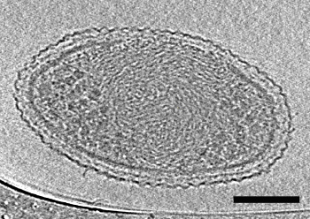
Cryo-electron tomograph of an ultra-small bacteria cell.
The scale bar is 100 nm.
(Lawrence Berkeley National Laboratory image.)
Jill Banfield, an
author of the study and a
professor in UC Berkeley's department of
Earth and Planetary Science, says that “we don't know the function of half the
genes we found in the organisms from these three phyla."[2] One interesting finding was that some of the bacteria have thread-like appendages. These so-called pili seem to enable inter-organism connections that aid in cellular processes.[2] This might be essential to their survival, since the limited genome would lack many functions.[1-2]
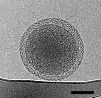
Cryo-transmission electron microscope image of an ultra-small bacteria cell showing pili-like structures at the surface.
The scale bar is 100 nm.
(Lawrence Berkeley National Laboratory image.)
As Banfield explains,
"These newly described ultra-small bacteria are an example of a subset of the microbial life on earth that we know almost nothing about... They're enigmatic. These bacteria are detected in many environments and they probably play important roles in microbial communities and ecosystems. But we don't yet fully understand what these ultra-small bacteria do."[2]
Birgit Luef of the
Norwegian University of Science and Technology (Trondheim, Norway), a former
postdoc in Banfield’s group, and an author of the paper, says that "there isn't a consensus over how small a free-living organism can be, and what the space optimization strategies may be for a cell at the lower size limit for life."[2]
This research was supported by the
U.S. Department of Energy.[2]
References:
- Birgit Luef, Kyle R. Frischkorn, Kelly C. Wrighton, Hoi-Ying N. Holman, Giovanni Birarda, Brian C. Thomas, Andrea Singh, Kenneth H. Williams, Cristina E. Siegerist, Susannah G. Tringe, Kenneth H. Downing, Luis R. Comolli, and Jillian F. Banfield, "Diverse uncultivated ultra-small bacterial cells in groundwater," Nature Communications, vol. 6, article no. 6372 (February 27, 2015 ), doi:10.1038/ncomms7372.
- Dan Krotz, First Detailed Microscopy Evidence of Bacteria at the Lower Size Limit of Life, Lawrence Berkeley Laboratory Press Release, February 27, 2015.
Permanent Link to this article
Linked Keywords: Audiophile; magnet; Alnico; magnetic moment; magnetic strength; rare-earth magnet; loudspeaker; energy conversion efficiency; inefficient; watt; electric power; amplifiers; high impedance; vacuum tube; transformer; magnetic core; frequency; 6L6; guitar amplifier; Wikimedia Commons; electronics; transistor; 14 nanometer process; International Technology Roadmap for Semiconductors; atom; silicon; nanometer; field effect transistor; material; graphene; science fiction; fan; positronic brain; computer scientist; benchmark; computational speed; multi-core processor; multiple processors; Advanced Micro Devices, Inc.; Opteron processor; symmetry; life; living; cell; virus; RNA; DNA; protein; self-replication; reproduction; reproductive mechanism; vacuum; scientist; University of California (Berkeley, California); Lawrence Berkeley National Laboratory (Berkeley, California); Joint Genome Institute (Walnut Creek, California); groundwater; electron microscope; electron microscopy; genome; genomic; volume; micrometer; Escherichia coli; human hair; filtration; filter; Rifle, Colorado; sterilization; sterilize; water; flash freezing; flash frozen; laboratory; cryogenic; transmission electron microscope; cytokinesis; dividing cell; whole genome sequencing; base pair; Archaea; phylum; phyla; spiral; ribosome; appendage; photograph; cell wall; cryo-electron tomograph; Jill Banfield; author; professor; Earth and Planetary Science; gene; environment; ecosystem; Birgit Luef; Norwegian University of Science and Technology (Trondheim, Norway); postdoctoral research; postdoc; United States Department of Energy.